Blood Vessels Labeled Brain : Bio Geo Nerd: September 2014 - Label the blood vessels of the male pelvis using the hints provided.. After ascending to the brain, the internal carotid arteries split into the anterior and middle cerebral arteries. The dense tight junctions between endothelial cells prevent paracellular transport through the. Sudden interruption of blood flow and oxygen to an area of brain tissue, which then may die (cerebrovascular accident, or cva, is another name for stroke.) ischemic stroke: Blood vessels are vital for the body and play a key role in diabetes helping to transport glucose and insulin. In the article on the ventricles within the cns, we will discuss their structure and.
Identify all of the blood vessels that are illustrated in the figure as you can while holding or otherwise examining whole brain specimens. Between arteries and veins, there is a network of. Blood vessels involved in small strokes. After ascending to the brain, the internal carotid arteries split into the anterior and middle cerebral arteries. This vessel supplies blood to the front part of your brain, knows as your frontal lobe.
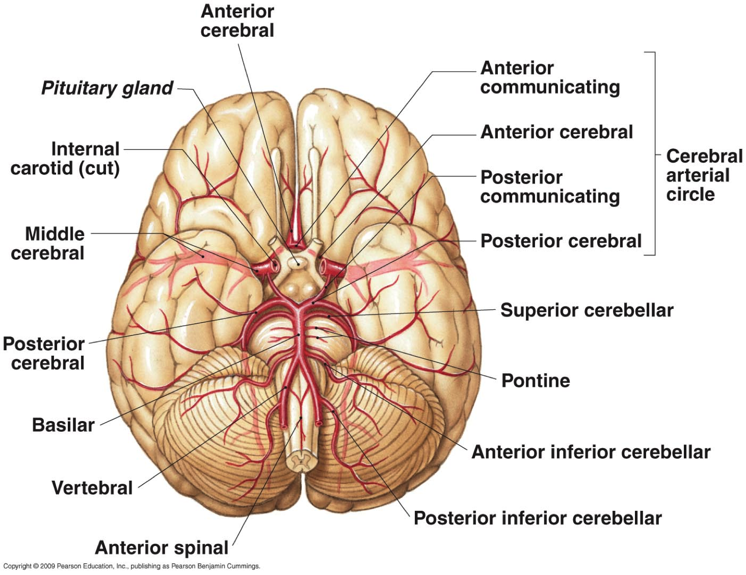
Role of TEM5 protein in brain blood vessel growth ... from www.sciguru.org A hemorrhage can result from a ruptured blood vessel, bleeding of an infarct, called hemorrhagic transformation, or from cancer that spread to the brain. There is a right sided aca and a left sided aca. Blood vessels in red in close communication with proliferating neuronal cells in the mouse cortex at embryonic day 10. Sudden interruption of blood flow and oxygen to an area of brain tissue, which then may die (cerebrovascular accident, or cva, is another name for stroke.) ischemic stroke: The two cell types ensure the integrity of the neural vasculature by maintaining the blood. Posterior communicating a internal carotid а. Label the veins of the anterior forearm and hand. Internal carotid artery (anterior circulation), vertebral artery (posterior circulation), and their hexagonal anastomotic network called blood brain barrier refers to the wall between the brain tissue and blood vessels.
Label heart and blood vessels.
In the condition known as cavernoma, lesions arise in a cluster of blood vessels in the brain, spinal cord or retina. Traditionally, pais has been explained as being caused by a blood clot forming within the ageing placenta, entering the fetal circulation, embolising across the patent foramen ovale, travelling into the left ventricle, into the ascending aorta and then one of the main three branches of the thoracic aorta. Red indicates arteries, and blue. The precise relation between blood vessels and brain regions, reflecting the physiology and pathology of brain function directly and accurately, has figure 3. Between arteries and veins, there is a network of. All blood vessels have some features in common. In the article on the ventricles within the cns, we will discuss their structure and. Only some of the vessels that exist in a real brain have been labeled. Blood vessels involved in small strokes. Blood vessels can be damaged by the effects of high blood glucose levels and this can in turn cause damage to organs, such as the heart and eyes, if significant blood vessel damage is sustained. The dense tight junctions between endothelial cells prevent paracellular transport through the. • identification of blood vessels as arteries, capillaries or veins from the structure of their walls. Blood travels from the heart in arteries, which branch into smaller and smaller vessels, eventually becoming arterioles.
Blood travels from the heart in arteries, which branch into smaller and smaller vessels, eventually becoming arterioles. The blood vessels (and nerves) enter the brain through holes in the skull called foramina. Another whole article within the blood vessels and csf section is dedicated to the cavernous sinus. Function and homeostasis of the brain relies on communication between its complex network of cells. Endothelial cells are labeled in red and pericytes in green.
Introduction to Neuroanatomy - Physiopedia from www.physio-pedia.com The anterior cerebral arteries supply the medial frontal and parietal lobes, and they are this interconnection can allow blood flow to continue if a major vessel is blocked on one side of the brain. While most blood vessels are located deep from the surface and. Blood vessels are referred to collectively as the vascular system and, together with the heart, make up the circulatory system or cardiovascular system. The carotid arteries and the vertebral arteries anterior cerebral artery (aca): After ascending to the brain, the internal carotid arteries split into the anterior and middle cerebral arteries. The blood vessels are the components of the circulatory system that transport blood throughout the human body. This new knowledge of the condition creates potential for developing better therapies for patients. • identification of blood vessels as arteries, capillaries or veins from the structure of their walls.
Red indicates arteries, and blue.
The walls of the arteries and veins both have the same. Blood vessel endothelium is continuous with the inner tissue lining of organs such as the brain, lungs, skin, and heart. Posterior communicating a internal carotid а. The two cell types ensure the integrity of the neural vasculature by maintaining the blood. Label the blood vessels in the inferior view of the brain using the hints provided. The carotid arteries and the vertebral arteries anterior cerebral artery (aca): Between arteries and veins, there is a network of. Cerebral arterial circle anterior communicating posterior cerebral a middle cerebral al reset zoom. This vessel supplies blood to the front part of your brain, knows as your frontal lobe. • identification of blood vessels as arteries, capillaries or veins from the structure of their walls. Blood vessels involved in small strokes. Fill in the blanks with the appropriate words to describe blood flow from the heart. Microscopically, it is formed by the endothelium of the blood vessel.
They also take waste and carbon dioxide away from the tissues. Fill in the blanks with the appropriate words to describe blood flow from the heart. Researchers can now show, at molecular level, that these changes originate in vein cells. The dense tight junctions between endothelial cells prevent paracellular transport through the. This is particularly important structure due to its clinical implications, which are discussed in more detail in the article.
THE BRAIN AND ITS BLOOD VESSELS | The Science Thinkers from www.thesciencethinkers.com Examine a second specimen and notice any differences, such as asymmetries in the size of the vertebral or posterior communicating arteries. In the article on the ventricles within the cns, we will discuss their structure and. Between arteries and veins, there is a network of. Blood in the brain is supplied by two pairs of large blood vessels (arteries): The anterior cerebral arteries supply the medial frontal and parietal lobes, and they are this interconnection can allow blood flow to continue if a major vessel is blocked on one side of the brain. Blood vessels can be damaged by the effects of high blood glucose levels and this can in turn cause damage to organs, such as the heart and eyes, if significant blood vessel damage is sustained. However, they have observed blood vessel damage caused. Fill in the blanks with the appropriate words to describe blood flow from the heart.
Blood vessels can be damaged by the effects of high blood glucose levels and this can in turn cause damage to organs, such as the heart and eyes, if significant blood vessel damage is sustained.
Examine a second specimen and notice any differences, such as asymmetries in the size of the vertebral or posterior communicating arteries. Sudden interruption of blood flow and oxygen to an area of brain tissue, which then may die (cerebrovascular accident, or cva, is another name for stroke.) ischemic stroke: • identification of blood vessels as arteries, capillaries or veins from the structure of their walls. Blood vessel endothelium is continuous with the inner tissue lining of organs such as the brain, lungs, skin, and heart. The walls of the arteries and veins both have the same. Blood vessels are vital for the body and play a key role in diabetes helping to transport glucose and insulin. Blood vessels in red in close communication with proliferating neuronal cells in the mouse cortex at embryonic day 10. These vessels transport blood cells, nutrients, and oxygen to the tissues of the body. Posterior communicating a internal carotid а. Blood in the brain is supplied by two pairs of large blood vessels (arteries): While most blood vessels are located deep from the surface and. Identify all of the blood vessels that are illustrated in the figure as you can while holding or otherwise examining whole brain specimens. After ascending to the brain, the internal carotid arteries split into the anterior and middle cerebral arteries.
This vessel supplies blood to the front part of your brain, knows as your frontal lobe blood vessels labeled. While most blood vessels are located deep from the surface and.


0 Komentar